Share this
by Neoteryx Microsampling on Jan 23, 2023 9:00:00 AM
A few years ago, Australian researchers Professor Tarl Prow and Li Lin, PhD, started working with Professor H. Peter Soyer and Dr. Alex Ansaldo at the University of Queensland (UQ) Dermatology Research Centre to develop a Microbiopsy device. According to an interview Professor Prow gave to Impact, a free-access series of reports on the impact of new scientific research, this new microsampling technology was intended to:
- Enable pain-free skin biopsy and blood collection
- Minimize the need for painful sampling procedures in the diagnosis of skin conditions and diseases
- Provide rapid diagnosis of skin issues, thus providing actionable information for faster treatment and recovery; and
- Move biopsy sampling out of medical facilities and into the field, thus helping those in remote geographical areas offer their study subjects and patients convenient diagnosis and treatment of skin conditions, including skin cancers and infectious skin diseases.
Here are highlights from the Q&A with Professor Prow in Impact, curated for readers of the Microsampling Blog at Neoteryx.com.
Q: Why this Microbiopsy device and why now?
A: The increase in incidents of skin disorders is leading to a corresponding increase in biopsies to extract sufficient material for analysis and diagnosis. Exciting new research into sub-millimeter extraction tools is helping to eliminate the need for painful and invasive extraction techniques.
Q: What are the sampling issues that you and your co-inventors were aiming to address?
A: Biopsies of suspect areas are now a routine process, but the process remains uncomfortable, risky, and difficult to carry out in non-medical facilities. Sample collection in the field is also of growing importance, but with the current methods this can be problematic. These challenges have led to an interest in micro-dermatological sampling tools that are minimally invasive and allow sampling under different conditions.
Traditional skin biopsies can involve harvesting a section of skin using either a sharp-edged tool such as a razor blade or a circular punch, which can vary in size from around 1.5mm up to 8mm in diameter, depending on the size of the sample required. Both methods usually result in some bleeding and may require one or more stitches to close them. Local anesthetic may be required, and post-procedure pain relief is often needed as well.
After wound healing, there may be a scar. Scarring can become an issue if several biopsies are required. This can lead to additional weeks of healing and unsightly results.
At the Future Industries Institute, University of South Australia and Dermatology Research Centre at The University of Queensland, Australia, we wanted to investigate a means of obtaining smaller, less invasive samples that don’t have such added complications.
Q: Where do you think your Microbiopsy technology will have the most benefit or impact?
A: We believe this Microbiopsy technology will be most useful in the form of an inexpensive device that can be used by general health professionals in clinics for skin disease testing, as well as in dermatology and cosmetology programs or studies that involve longitudinal testing. The device is designed for cosmetically sensitive areas and would be ideal for use on children or people with many lesions.
Q: Can you describe how Microbiopsies work?
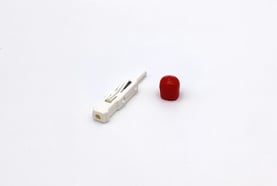 A: Our Microbiopsy device has a modified spring-loaded blood lancet applicator. When applied, the microneedle captures a tiny sample of blood and skin. This sub-millimeter Microbiopsy platform technology is less painful than a conventional blood lancet, so there is no need for local anesthetics. It has a safety profile similar to existing blood lancets.
Q: What are the advantages of microsampling with this Microbiopsy technology over traditional harvesting techniques?
A: Conventional methods of obtaining skin specimens involve invasive surgical techniques such as shave biopsy or Mohs surgery that require local anesthetic injection and sutures that can lead to scars. Microsampling is quick and relatively painless, which means researchers or physicians can sample many more areas than conventional biopsy approaches and repeat sampling over time.
Conventional biopsy specimens are then preserved with formalin to maintain structural organization and sent for histopathological analysis. Histopathological sections are rarely used for molecular fingerprinting in dermatology. Microsampling using our Microbiopsy platform not only provides samples that are of higher quality than formalin fixed histopathological samples, but also have the capacity to target specific areas within a lesion.
Q: How will this technology benefit other areas of dermatology research?
.jpg?width=157&height=209&name=Microbiopsy%20IM_3821_RGB_1024px_72dpi%20(1).jpg)
A: Surprisingly, one area of great potential benefit is the cosmetics field. Their only options have been cell culture research, non-invasive analysis and volunteers undergoing conventional biopsies. Now they are testing cosmetics in volunteers and taking Microbiopsies to evaluate molecular changes in the treated skin. This can be a very powerful device for cosmeceutical development and testing.
We are now working with groups around the globe who each have their own areas of investigative dermatology where Microbiopsy is playing a large role. In Munich, Germany, for example, we are working with researchers learning about infant inflammatory disease. In Israel we have collaborators who are using Microbiopsy samples to detect leishmania in volunteers from Ghana and Ethiopia. In Brazil, our collaborators are applying Microbiopsies to dogs to investigate signs of parasitic infection. In New York, USA, our collaborators are using Microbiopsies to examine the molecular landscape of suspicious nevi in patients at risk for melanoma. Together we have performed over 2,200 Microbiopsies on many different body sites, including the face and neck.
Q: Do you have anything further you’d like to add about the Harpera Microbiopsy Punch?
A: Harpera uniquely enables doctors and researchers to take tiny skin samples from infants and children in addition to all body sites, including the face. This allows researchers to make new discoveries that will lead to rapid pediatric healthcare advances not possible before. Likewise, being able to take tiny samples from the face will fuel research in healthy ageing and lead to improvements in diseases that affect the face. At the heart of this is that Harpera enables researchers to move away from animal models that are not directly relevant to studying human skin diseases.
We are learning that there are many uses for this technology that we never imagined when we first developed the idea. We look forward to seeing how we can help these innovators put our technology to work!

This is curated content, which was summarized for our readers. To learn more about the important research outlined in this blog, visit the original article that appeared in Impact.
Image Credits: iStock, Neoteryx, Trajan
In 2017, the co-inventors at UQ entered into an evaluation partnership with Trajan Scientific and Medical, a global developer and manufacturer of analytical and life sciences products and tools headquartered in Melbourne, Australia. That partnership led to a new license agreement in 2022, whereby Trajan agreed to further develop the UQ team’s microbiopsy technology for commercialization through Neoteryx, its microsampling brand. The technology has been named the Harpera™ tool.
Since collaborating on the microbiopsy tool that has now become Harpera, the inventors from UQ have been involved in other interesting endeavors in the field of dermatology. Professors Soyer, Ansaldo and Li continue their work in Australia, while Professor Prow now works at Hull York Medical School in the United Kingdom, where he has recently taken on the role of Director of their Skin Research Centre. You can learn more about his work via this TED Talk he gave in 2019.
For more information, please visit the Harpera page on the Neoteryx website. If you are interested in applying this tool, please contact us at: info@neoteryx.com.
Related Reading:
Prow, T.W., et al. (2013). "The opportunity for microbiopsies for skin cancer." Future Oncol9 (9): 1241-1243
Dermatology Research Centre, School of Medicine, The University of Queensland (March2016), “Natural history and properties of naevi in advanced melanoma patients receiving treatment”, ACTRN 12616000272493 (ANZCTR)
Lin, L .L. and T.W. Prow (2017). "Novel microdevices for controlled blood and skin extraction, NHMRC." Impact 2017(6): 58-60.
Lin, L.L.,et al. (2013). "Microbiopsy engineered for minimally invasive and suture-free sub-millimetre skin sampling." F1000Res 2: 120.

No Comments Yet
Let us know what you think